L3 Cell Biology
🧠 Concepts Introduced
LIST
FROM "00 Archive/Objects/Concepts"
WHERE lecture = this.file.link
Learning Objectives
- Describe the chemical composition and functions of membrane
- Explain the importance of membrane fluidity
- Understand the principle in the movement of substances across cell membranes
- Distinguish different types of membrane proteins in membrane transport
Cell Membrane
The cell membrane is composed of three main types of components by mass: Lipids (40%) Proteins (50%) Carbohydrate portions of glycolipids and glycoproteins (10%) (act as receptor)

Cellular Membrane Functions
-
Compartmentalization/protection: separating molecules and ions between intra-cellular/-organellar and extra-cellular/-organellar spaces
-
Providing selectively permeable barrier for import and export of molecules and ions: Not all solutes can pass through membrane freely
-
Cell communication: Intercellular interaction, responding to external stimuli
-
Cell growth and motility
-
Energy generation (will be discussed in next lecture: METABOLISM)
1. Compartmentalization
Membranes form "compartments" in the form of organelles Why having different organelles?
Each organelle is a discrete unit, its enclosed by a membrane, and has a specialized function, eg. the nuclear membrane separate transcription and translation in eukaryotes. Enzymes and substrates are not spread throughout the entire cytoplasm
Folded membrane increases the surface areas and the capacity of production, eg. energy-producing reaction happens at the folded surface of the inner membrane of mitochondria.

2. Selective Permeability Barrier

charged ions are hydrophobic
3. Cell Communication
Signal Transduction: Proteins shaped into receptors embedded in the cell's membrane. Once a ligand binds to its receptor, the signal is transmitted through the membrane and into the cytoplasm.
trigger signal downstream. cascading down

Extracellular Vesicles: lipid bound vesicles secreted by cells into the extracellular space. Lipids, nucleic acids, and proteins can be found inside the vesicle.

Cell Junction: Gap junctions are specialized connections that form a narrow pore between adjacent cells. These pores permit small molecules and ions to move from one cell to another. (explained in Connexon)

4. Cell Movement
Maintaining cell structure via its interaction with the cytoskeleton & ECM Cell movements are mediated by integrin attachment
 extracellular matrix: contain carbohydrates and proteins
Plasma membrane & Phospholipids
Lipid Bilayer
Phospholipid bilayer is less than 10 nm thick.
Amphipathic: each lipid has a hydrophilic head (water soluble) and a hydrophobic tail (water insoluble)
This design makes the self-sealing form of the lipid bilayer
Lipid bilayer is flexible and fluidic (membrane fluidity)
Hydrophilic head: either charged or uncharged polar groups that form electrostatic attractions or hydrogen bonds with water molecules
Hydrophobic tail: uncharged and nonpolar (aggregate with other hydrophobic molecules)
choline can be serine here as well

double bond between two carbon forms the bend = kink
Types of phospholipids
Four major phospholipids in mammalian plasma membrane: phosphatidylethanolamine (PE), phosphatidylserine (PS), phosphatidylcholine (PC) and sphingomyelin (SM).
sphingomyelin is important important as it appears in a lot of cell type toxin attach to it as it appears a lot
PE, PS and PC are phosphoglycerides, which have 3-carbon glycerol backbone.

Phosphoglyceride - simple form of phospholipid Two carbons are linked to fatty acid chains with ester bonds and one is attached to the phosphate group.
Sphingosine - simple form of sphingolipid Sphingomyelin is built from sphingosine which has (1) a long hydrocarbon chain with one double bond; (2) an amino (NH2) group on carbon 2; (3) two hydroxyl (OH) groups on carbons 1 and 3; (4) choline as polar head group
Membrane Fluidity
Core lipid bilayer exists in a fluid state, capable of movement
Individual lipid molecules move within their monolayer, hence lipid membranes are highly asymmetric.
Motion of individual lipid molecules includes lateral diffusion, rotation and flexion
Transverse diffusion of single lipid molecule is rare
 flexion - moving of the tail
flip flop - one layer to another layer
Length and number of double bonds affect how tightly they pack in the bilayer
Shorter fatty acid chains increase the fluidity of bilayer
The chain with double bond (unsaturated) fatty acid creates a small kink in the tail, making the bilayer more fluid The kink gives it space between phospholipid for it to move around
- Membrane fluidity depends on its temperature and its composition (saturated vs unsaturated).

- The temperature at which a membrane solidifies depends on the types of lipids
- More importantly, the physical state of the membrane is affected by cholesterol
high temperature - more saturated fatty acids. move too much it will break low temperature - less saturated, fatty acids more kink
Lipid Bilayer - Cholesterol
Cholesterol is present in large amounts (20% of lipids) in the plasma membrane They are short and rigid, filling up spaces between phospholipid left by kinks
It stiffens the bilayer, making it less flexible and less permeable
Cholesterol can be considered a fluidity buffer for the membrane, resisting changes in membrane fluidity:
- restrains phospholipid movement at warm temperatures, and
- prevents tight packing at cool temperatures


Importance of Membrane Fluidity
-
Membrane proteins can rapidly move to interact with each other, which is essential in cell signalling
-
Permit membrane proteins and lipids to leave cell membrane to other sites
-
Ensure that membrane molecules are distributed evenly between daughter cells during cell division
-
Allow fusion of cell membrane (membrane assembly) - endocytosis and exocytosis
-
Facilitate cell adhesion and migration
1. Membrane proteins can rapidly move to interact with each other, which is essential in cell signalling
Membrane proteins form a mosaic of particles penetrating the lipid bilayer
Amphipathic: hydrophobic part(s) of membrane protein interact with lipid bilayer and hydrophilic part(s) are exposed to either extracellular space or/and cytosol.
Two major types of membrane protein: integral and peripheral membrane proteins
lon transporter and ligand receptor are examples of integral and peripheral membrane proteins, respectively.


2. Permit membrane proteins and lipids to leave cell membrane to other sites
Extracellular vesicles (EV) serve as vehicles for cell-to-cell communication through horizontal transfer of bioactive molecules (proteins, lipids and nucleic acids).
EVs produced from a secreting cell may be internalized by fusion (1), endocytosis (2), phagocytosis (3) or may interact with the membrane proteins of the target cell (4).

3. Ensure that membrane molecules are distributed evenly between daughter cells during cell division
Plasma membrane can only be replicated from the existing membrane
The Golgi apparatus is disassembled during prometaphase into vesicles, which are segregated in metaphase and then reformed during telophase.
Mitochondria are fused in interphase, fragmentated during mitotic entry in prophase and begin to re-fuse during anaphase

- The Principle of Membrane Continuity
- "Plasma membrane can only be replicated from the existing membrane."
- Just as cells only arise from pre-existing cells (omnis cellula e cellula), biological membranes grow exclusively by expansion and remodeling of pre-existing membranes.
- During cell division, the cell must carefully expand its surface area and distribute its membrane molecules (lipids, integral proteins, and peripheral proteins) evenly between the two emerging daughter cells during cytokinesis.
- Golgi Apparatus Cycle (Disassembly and Reformation) As shown in the diagram, the Golgi apparatus undergoes a dramatic structural cycle to ensure it is partitioned correctly:
- Prometaphase (Disassembly): The Golgi breaks down into small vesicular clusters and tubules. This prevents it from getting tangled in or blocking the mitotic spindle.
- Metaphase (Segregation): These dispersed vesicles are segregated and scattered across the cell, ensuring they are present on both sides of the dividing cell.
- Telophase (Reformation): Once chromosomes reach the poles and the nuclear envelope reforms, these vesicles fuse back together to rebuild intact, functional Golgi stacks in each daughter cell.
- Mitochondrial Dynamics (Fusion and Fragmentation) Mitochondria are dynamic organelles that constantly change shape based on the cell cycle:
- Interphase (Fusion): During normal cellular metabolism and growth, mitochondria exist as long, interconnected, fused networks that maximize energy production and share mitochondrial DNA.
- Prophase (Fragmentation): As the cell enters mitosis (prophase), these large networks fragment into numerous smaller, individual mitochondrial units. This fragmentation is crucial because, unlike a single large network, small mitochondria can be easily and evenly distributed across the dividing cytoplasm into both daughter cells.
- Anaphase/Telophase (Re-fusion): Once cell division is underway, the mitochondria begin to re-fuse back into extended networks to restore normal energy homeostasis in the new daughter cells.
4. Allow fusion of cell membrane (membrane assembly) - endocytosis and exocytosis

explained in Receptor-mediated endocytosis

5. Facilitate cell adhesion and migration
Cell migration is the normal movement of cells from one place to the other in response to chemical signals.
Cell migration is involved in embryonic development, cell differentiation, tissue regeneration, wound healing, responding for immune signals, cancer metastasis, etc.
- Cell invasion is a type of aberrant cell migration which is related to cancer.
- Cells migrate through the extracellular matrices and penetrate into tissues or infiltrate into neighbouring new tissues.
cells have to move to particular position in embryonic development cell differentiation on cell migration
Membrane Transport
Membrane transport functions
-
Maintain the cell function allows cells to bring in oxygen and nutrients (glucose, polysaccharides, amino acids, steroids) and release waste
-
Maintain homeostasis (ie. interaction with the environment)
-
Plasma membrane is selectively permeable
Selective Permeability of Lipid Bilayer
Plasma membrane is amphipathic, containing hydrophilic and hydrophobic regions
Speed of molecules to pass the membrane depends on size (large or small) and solubility (polar or nonpolar; charged or uncharged).
In general, the smaller, the more hydrophobic or nonpolar the molecule is, the faster it will diffuse across the membrane.
Small nonpolar and small uncharged polar molecules dissolve readily in lipid bilayers and diffuse across them Large uncharged polar molecules such as glucose can hardly cross the membrane Charged ions are impermeable no matter how small they are, due to their strong electrical attraction to water
 water soluble or polar solutes pass through the membrane need to travel with the help of membrane transport proteins
Types of Membrane Transport

Passive Transport
Simple diffusion: solutes or molecules (gases, steroid) travel from high to low concentration gradient across membrane
Facilitated diffusion: transport facilitated by specialized membrane transport proteins
Osmosis: water moves from a region of higher water concentration (lower solute concentration) to one of lower water concentration (higher solute concentration)
Osmosis (water diffusion)
Tonicity is the ability of a solution to cause a cell to gain or lose water
Isotonic solution: Solute concentration is the same as that inside the cell; no net water movement across the plasma membrane
Hypertonic solution: Solute concentration is greater than that inside the cell; cell loses water
Hypotonic solution: Solute concentration is less than that inside the cell; cell gains water
Osmosis (plant)
Diffusion of water in plant cells Plant cells develop turgor in hypotonic solutions because cell walls prevent swelling.
If the cell and its surroundings are isotonic, there is no net movement of water into the cell: the cell becomes flaccid (limp), and the plant may wilt
In hypertonic solutions the plant cell undergoes plasmolysis and dies
Aquaporins
Water are small, uncharged, polar molecules, they can travel across the phospholipid bilayer via
- Simple diffusion (but the diffusion rate is slow). Aquaporin (water channel)
- Aquaporins are specialized channel proteins formed as a tetramer, each subunit has six membrane-spanning alpha helical domains
Found in all living organisms (bacteria, archaea, plants and animals)
In kidney, the presence of aquaporins in the epithelium of distal, Cell collecting tubules help to uptake water - a process of reabsorption of water.
entire aquaporins has 4 subunit which builds a tube

Facilitated Diffusion

Membrane Proteins & Functions Membrane proteins are divided into TWO main classes (different in terms of function)
Channels (passive transport)
- form tiny hydrophilic pores across membrane to allow substances (usually
- inorganic ions) to pass by diffusion such as ion channels Allow any ion or molecule that is small and carries appropriate charge to pass through
Transporters
- shift small organic molecules or inorganic ions from one side of the membrane to another by changing shape
- Transfer only molecules that fit into specific binding sites on the proteins (similar to how enzyme-substrate system works)

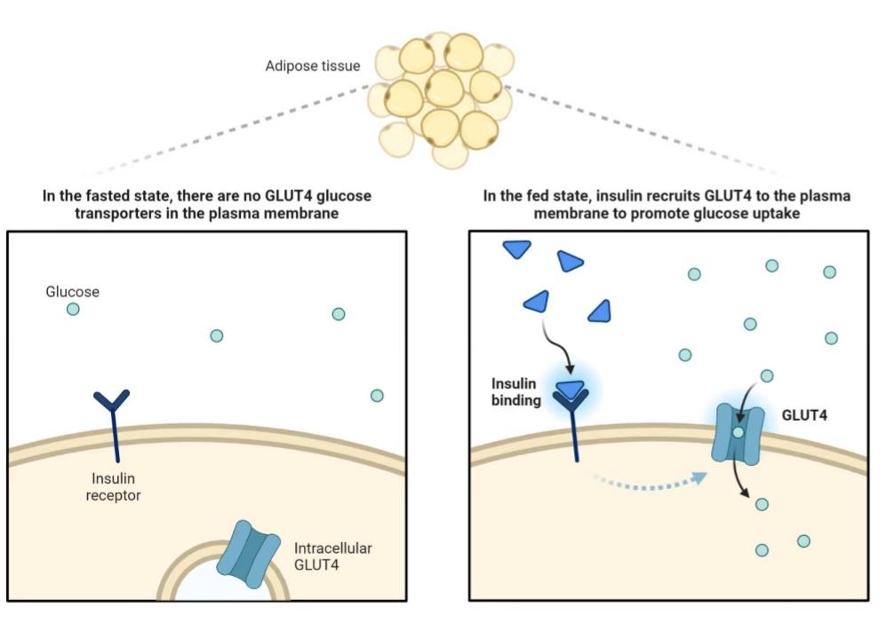
Facilitated Diffusion: GLUT4 Transporter Responsible for transportation of glucose molecules Highly selective. eg. glucose transporter (GLUT4). the binding sites in the glucose transporter bind only D-glucose but not its mirror image, L-glucose which cannot be used for glycolysis
- Blood sugar level is HIGH
- Insulin hormone are released (by endocrine cells of pancreas) and binds to its receptor
- Triggers GLUT4-containing vesicles moving to the cell surface
- Stimulates uptake of glucose into various tissues (skeletal muscles, fat cells)
Transporters Transporters can function as uniports, symports or antiports Uniports - carry single solute across membrane Coupled transporters - move multiple solutes
- Symports - transfer solutes in the same direction
- Antiports - transfer solutes in the opposite direction Secondary active transport
All of these transporters can be used for either passive or active transport
Active Transport
Facilitated diffusion is passive because the solute moves down its concentration gradient
Some transport proteins can move solutes against their concentration gradients → Active transporters
Active transporter or pumps require energy in different forms:
- ATP (ATP-driven pumps): Primary Active Transport
- Absorbance of light (light-driven pumps)
- Flow of other substances down their gradient: Secondary Active Transport or Co-transporter
Na+/K+ ATPase Pump (Active Transport)
The Na+ K+ ATPase pumps 3 Na+ out of the cell and 2 K+ that into the cell, for every single ATP consumed.
Pump has higher affinity to Na+ than K+, it binds ATP and 3 Na+, leading to conformational change.
Phosphorylated pump binds strongly to K+ than Na+, hence releasing Na+ extracellularly and binds 2 K+.
The Na+ K+-ATPase pump maintains the gradient of a higher concentration of sodium extracellularly and a higher level of potassium intracellularly.
It works to maintain the resting membrane potential of a neuron.
Co-transport
Gradients created by active ion pumping store energy that can be coupled to other transport processes.
Cotransport is also called secondary active transport
For example, the sodium-glucose cotransporter 1 (SGLT1) couples the transport of 2 Na+ ions moving down their gradient with the uptake of 1 glucose molecule against its gradient across the apical membrane of intestinal epithelial I cells cells and renal proximal tubule cells.
Proton pump moves hydrogen from low to high concentration gradient (require ATPs)
Symport lets hydrogen ions move down their gradient, but simultaneously brings a sucrose molecule up its gradient and into the cell
